

When it comes to diagnosing neurological disorders and conditions that affect the brain and nervous system, neurologists employ a wide array of diagnostic procedures and tools to unravel the complexities hidden within the human brain and nervous system. These procedures play a pivotal role in identifying and understanding various neurological conditions, enabling neurologists to provide targeted treatment and care. In this blog, we will delve into the fascinating world of neurology and explore the different types of diagnostic procedures that neurologists utilize. Whether you’re a patient seeking insight into your own neurological health or simply curious about the field of neurology, this exploration will shed light on the many ways that doctors diagnose and understand the enigmatic realm of the human brain. So, let’s embark on this enlightening journey and uncover the secrets behind the art and science of neurological diagnosis.
The Foundation of Neurological Diagnosis
Before we delve into the intricate diagnostic procedures employed by neurologists, it’s essential to understand the foundational elements of neurological diagnosis. Much like any medical specialty, the diagnostic process in neurology begins with a thorough medical history and physical examination. These initial steps are crucial as they allow neurologists to gather valuable information about a patient’s symptoms, medical background, and family history.
During the medical history assessment, patients are encouraged to describe their symptoms in detail, including when they began, their frequency, and any associated factors. Neurologists may inquire about lifestyle factors, such as diet and sleep patterns, that could be relevant to the patient’s condition. Additionally, a family history review helps identify any genetic predispositions to neurological disorders.
The physical examination in neurology is particularly focused on assessing the nervous system. Neurologists look for signs of neurological dysfunction, such as changes in muscle strength, reflexes, coordination, and sensation. Specialized tests like the Mini-Mental State Examination (MMSE) can assess cognitive function and detect signs of conditions like dementia. These initial steps set the stage for further diagnostic investigations, ensuring that the subsequent procedures are tailored to the patient’s specific needs.
Unraveling Mysteries with Neuroimaging
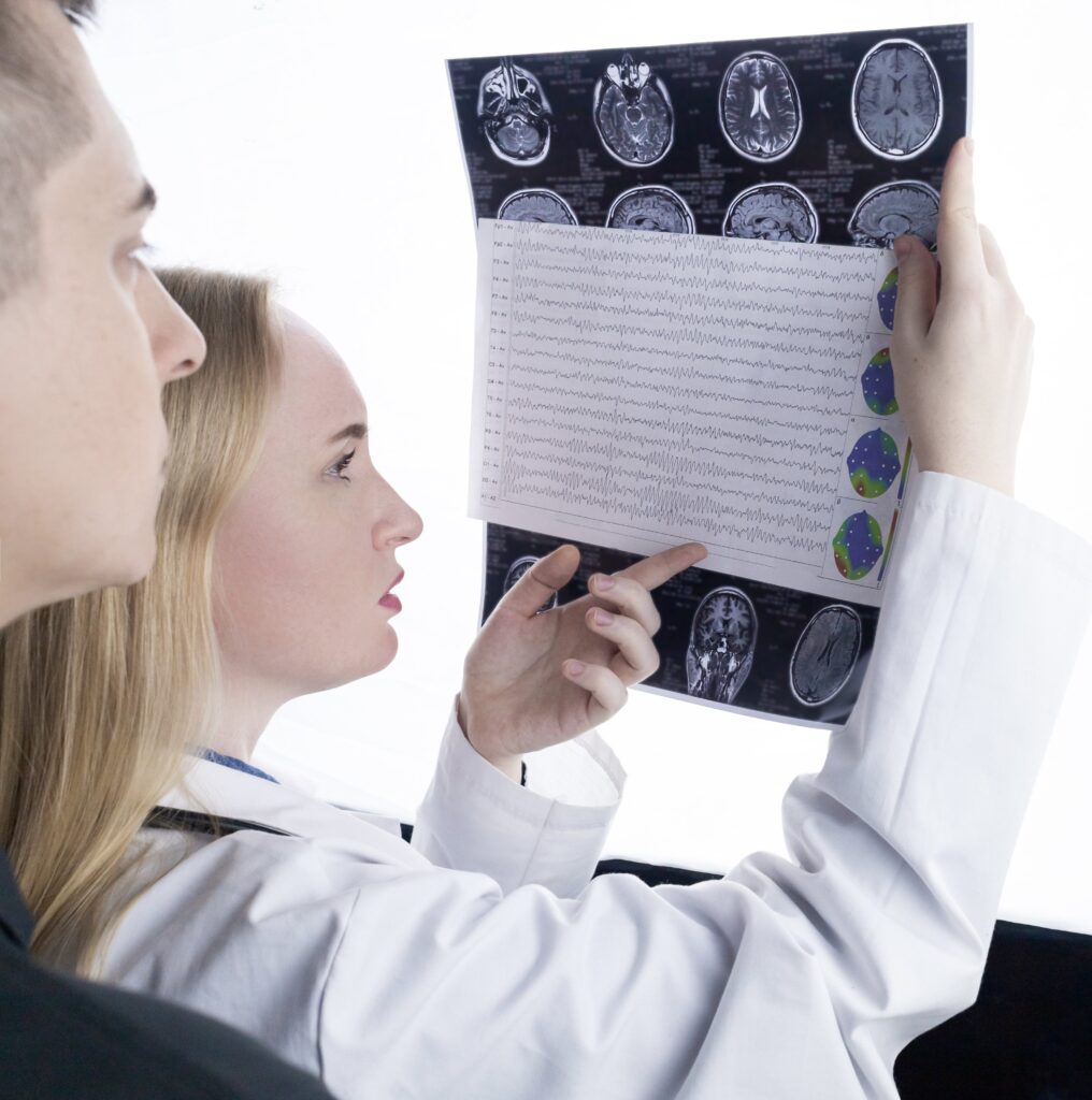
One of the most powerful diagnostic tools in the neurologist’s arsenal is neuroimaging. This technology allows doctors to peer inside the brain and visualize its structure and function. Magnetic Resonance Imaging (MRI) and Computed Tomography (CT) scans are commonly used to provide detailed images of the brain’s anatomy, helping identify abnormalities like tumors, strokes, or structural malformations. Additionally other neuroimaging techniques may be used such as:
- Positron Emission Tomography (PET)
- Single-Photon Emission Computed Tomography (SPECT)
- Diffusion Tensor Imaging (DTI)
- Magnetic Resonance Spectroscopy (MRS)
- Functional Near-Infrared Spectroscopy (fNIRS)
- Ultrasonography
- Cerebral Angiography
Functional imaging techniques, such as Positron Emission Tomography (PET) and Single-Photon Emission Computed Tomography (SPECT), go beyond structure to reveal how different parts of the brain function. These scans are vital in diagnosing conditions like epilepsy, Alzheimer’s disease, and movement disorders.
Electrophysiological Tests: Measuring Brain Activity
Neurologists use a variety of electrophysiological tests to assess the electrical activity of the nervous system. These tests help diagnose and monitor neurological conditions by measuring the transmission of electrical signals within nerves and muscles. Here are some of the common electrophysiological tests used by neurologists:
Electroencephalography (EEG):
EEG records the electrical activity of the brain by placing electrodes on the scalp. It is used to diagnose and monitor conditions such as epilepsy, seizures, and certain sleep disorders. EEG can detect abnormal patterns of brain activity and help guide treatment decisions. There are two types of EEG. An ambulatory EEG is a portable and long-term monitoring system that records brain activity over an extended period while a patient goes about their daily activities, whereas a traditional electroencephalography (EEG) is typically conducted in a clinical setting and records brain activity during a shorter, controlled session.
Nerve Conduction Studies (NCS):
NCS measures the speed and strength of electrical signals as they travel along peripheral nerves. Electrodes are placed on the skin, and a small electrical stimulus is applied to one part of the nerve, while the response is recorded from another part. NCS helps diagnose conditions like carpal tunnel syndrome, peripheral neuropathy, and nerve injuries.
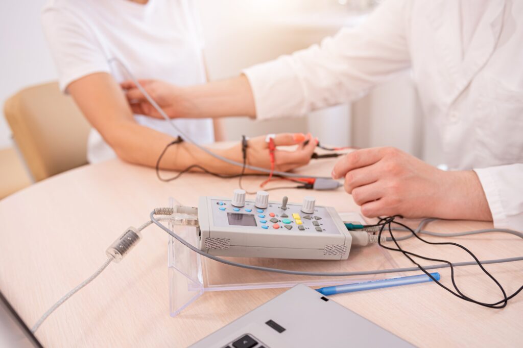
Electromyography (EMG):
Electromyography (EMG) assesses the electrical activity of muscles. A fine needle electrode is inserted into the muscle, and the electrical signals generated during muscle contraction and relaxation are recorded. EMG is valuable for diagnosing muscle disorders, motor neuron diseases, and nerve compression syndromes.
Evoked Potentials:
Evoked potentials tests measure the electrical response of the nervous system to specific stimuli, such as visual, auditory, or sensory stimuli. Common types of evoked potentials include Visual Evoked Potentials (VEP), Auditory Brainstem Responses (ABR), and Somatosensory Evoked Potentials (SSEP). Evoked potential testing helps diagnose conditions like multiple sclerosis and assess sensory and auditory pathways.
Neuropsychological Testing and Clinical Assessments
Neuropsychological testing is a specialized assessment process that involves the evaluation of an individual’s cognitive, emotional, behavioral, and psychological functioning. It is conducted by clinical neuropsychologists, who are trained to assess how various aspects of brain function may be impacting a person’s behavior and cognitive abilities.
Neuropsychological testing typically includes a battery of standardized tests and measures that assess a wide range of cognitive functions, including:
- Memory: Tests to evaluate short-term memory, long-term memory, and working memory.
- Attention and Concentration: Assessments of a person’s ability to sustain attention, switch attention between tasks, and resist distractions.
- Language: Evaluation of language skills, including comprehension, expression, and fluency.
- Executive Functioning: Tests of higher-level cognitive abilities, such as problem-solving, planning, organization, and decision-making.
- Visuospatial Skills: Assessment of an individual’s ability to perceive and interpret visual information and spatial relationships.
- Motor Skills: Evaluation of fine and gross motor skills, coordination, and dexterity.
- Emotional and Psychological Functioning: Assessment of emotional regulation, mood, and psychological well-being.
Neuropsychological testing is often used in clinical settings to help diagnose and understand various neurological and psychological conditions, including traumatic brain injuries, strokes, neurodegenerative diseases (e.g., Alzheimer’s disease), learning disorders, attention-deficit/hyperactivity disorder (ADHD), and psychiatric conditions.
The results of neuropsychological testing provide valuable insights into an individual’s cognitive strengths and weaknesses, which can guide treatment planning, rehabilitation, and interventions. Additionally, these assessments can help track changes in cognitive function over time and monitor the progression of neurological conditions.
Other Types of Testing
Vestibular Function Testing
Vestibular function testing is a specialized area of diagnostic testing focused on evaluating the function of the inner ear and the vestibular system, which plays a crucial role in balance and spatial orientation. A Videonystagmogram (VNG) is a diagnostic test that specifically assesses the function of the vestibular system by measuring eye movements, particularly nystagmus (involuntary rapid eye movements), in response to various stimuli. It is commonly used to diagnose and assess conditions related to balance and dizziness, such as benign paroxysmal positional vertigo (BPPV), Meniere’s disease, labyrinthitis, and other vestibular disorders.
Neuromuscular Ultrasound
Neuromuscular ultrasound is a diagnostic imaging technique that involves the use of high-frequency sound waves to create real-time images of muscles, nerves, and surrounding tissues. During a neuromuscular ultrasound examination, a transducer is placed on the skin’s surface, and sound waves are directed into the targeted area. These waves bounce back as echoes and are converted into detailed images that reveal the structure and function of muscles and nerves. Neurologists can utilize neuromuscular ultrasound to assess conditions such as peripheral neuropathy, muscle disorders, nerve compression syndromes like carpal tunnel syndrome, and to guide procedures like nerve blocks or injections.
Skin Biopsy
Small fiber neuropathy primarily affects the small nerve fibers responsible for pain and temperature sensation, and it can be challenging to diagnose through traditional neurological examinations and nerve conduction studies. Therefore, neurologists will sometimes perform a skin biopsy to diagnose small fiber neuropathy. Skin biopsy allows neurologists to directly examine these small nerve fibers in a minimally invasive manner. By analyzing a small skin sample, typically taken from the lower leg, under a microscope, neurologists can assess the density of small nerve fibers and identify abnormalities, such as reduced nerve fiber density or signs of damage. This diagnostic approach provides objective and quantifiable evidence of small fiber neuropathy, aiding in the confirmation of the condition and guiding appropriate treatment strategies to alleviate patients’ pain and discomfort.
Conclusion:
The field of neurology is a fascinating journey into the complexities of the human nervous system. By utilizing an array of diagnostic procedures ranging from traditional clinical assessments to cutting-edge technologies like neuroimaging and genetic testing, neurologists can unlock the mysteries of the brain and provide patients with the answers and care they need.

Dr. Kashouty, a diplomate of the American Board of Psychiatry and Neurology (ABPN), practices general neurology with fellowship trained specialization in clinical neurophysiology. Dr. Kashouty finds the form and function of the nerves and muscles the most interesting part of neurology, which is what led him to specialize in neurophysiology with more emphasis on neuromuscular conditions. He treats all neurological diseases, but his main focus is to treat and manage headaches, movement disorders and neuromuscular diseases.