
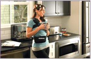
Ambulatory EEG
An ambulatory electroencephalogram (EEG) is a non-invasive test that records your brain activity for 49-96 hours during your usual daily routine. Prior to the test, you will have electrodes attached to your scalp using a temporary glue. During the test, these electrodes will monitor your brain’s electrical activity for your neurologist to evaluate…
Computerized Neuropsychological Evaluation
Braincheck Memory is a neurocognitive assessment developed to track and monitor changes in your cognitive health. It works by playing a series of neurocognitive games that are each designed to evaluate and measure a specific aspect of brain health. A cognitive care plan will then be developed based upon the information obtained…
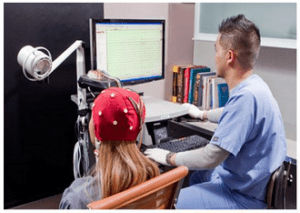
Electroencephalography EEG
Electroencephalography (EEG) is a non-invasive test that measures and records the brain’s electrical activity over a specified period of time. During an EEG, you will be positioned in a bed or chair and around 20 electrodes will be attached to your scalp to continuously record your brain activity. Most EEGs take about an hour, including preparation…
Evoked Potential Testing
There are three different types of evoked potential testing. The first type is Visual Evoked Potential (VEP) that evaluates the visual function of the brain by recording the optic nerve pathways. The second type is Brainstem Auditory Evoked Potential (BAEP) which evaluates brainstem function. Finally, the third type is Somatosensory Evoked Potential (SSEP) which evaluates the slowing of sensory nerve pathways…
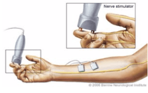
Nerve Conduction Study and EMG
Nerve conduction studies and needle electromyography (EMG) are two tests used to evaluate the peripheral nervous system. Nerve conduction studies are performed by placing surface electrodes along peripheral nerves. EMGs are performed by placing a small needle in the muscle to evaluate muscle and nerve functions…
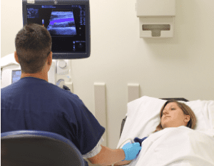
Neuromuscular Ultrasound
A neuromuscular ultrasound produces images of the peripheral nerves to allow physicians to identify and diagnose neuromuscular diseases. This diagnostic technique is painless, non-invasive, and cost-effective. Compared to other imaging methods, ultrasound produces high resolution images that are portable and use decreased amounts of radiation…
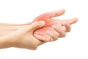
Skin Biopsy for Small Fiber Neuropathy
Since small fibers travel too slow, their conduction response cannot accurately be measured by nerve conduction studies or EMG tests. Therefore, taking a small skin sample from the ankle and below the hip allows your neurologist to determine whether damage to the unmyelinated fibers in the peripheral nerves has occurred…
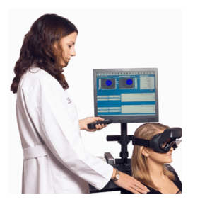
VNG (Videonystagmography)
Videonystagmography (VGN) technology is used to examine the inner ear and central motor functions to determine if inner ear disease is the cause of dizziness or balance problems. There are four parts of VGN testing including: ocular mobility, optokinetic nystagmus, positional nystagmus, and caloric testing…